Gunnison, CO
November 10, 2026
Heights Physical Therapy
700 N. Main St, Unit B
Gunnison, CO 81230
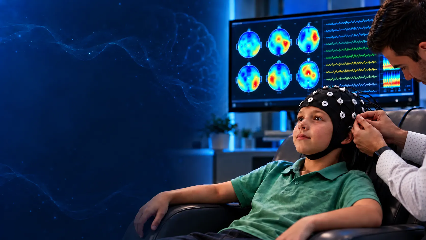
Two people can have the same symptoms, yet have very different patterns of brain activity causing them. A qEEG brain map allows us to objectively measure the brain’s electrical activity and see which areas may be overactive, underactive, or not communicating efficiently. Instead of guessing based on symptoms alone, we start by looking at the brain itself.
How the sensors work
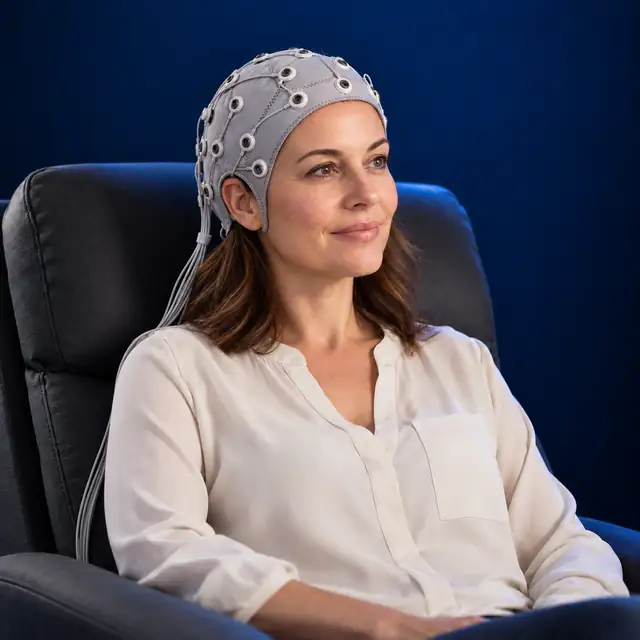
Your brain already produces tiny electrical signals. EEG sensors rest on your scalp and record those signals while you sit comfortably.
Groups of brain cells produce tiny electrical changes.
Scalp electrodes detect differences in these signals.
The recorder amplifies the signal and displays brainwaves.
Learn about EEG recording: NHS · Johns Hopkins Medicine
A brain map takes about 20 minutes while you sit in a comfortable chair. It is safe and non-invasive, and NO electrical activity is sent into your brain. It only records the electrical activity your brain naturally produces, similar to how an EKG records your heart’s electrical activity. We generally provide brain mapping for ages 5 to 100.
Objectively identify the patterns that need support.
Customized brain training to address issues on your initial brain map.
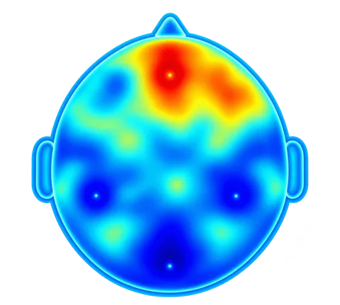
Objectively measure progress and refine your training.
Continue or adapt brain training based on your mid map progress.
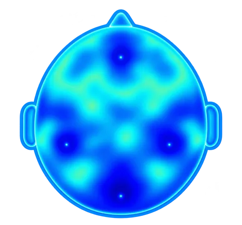
Objectively verify whether brain activity has normalized.
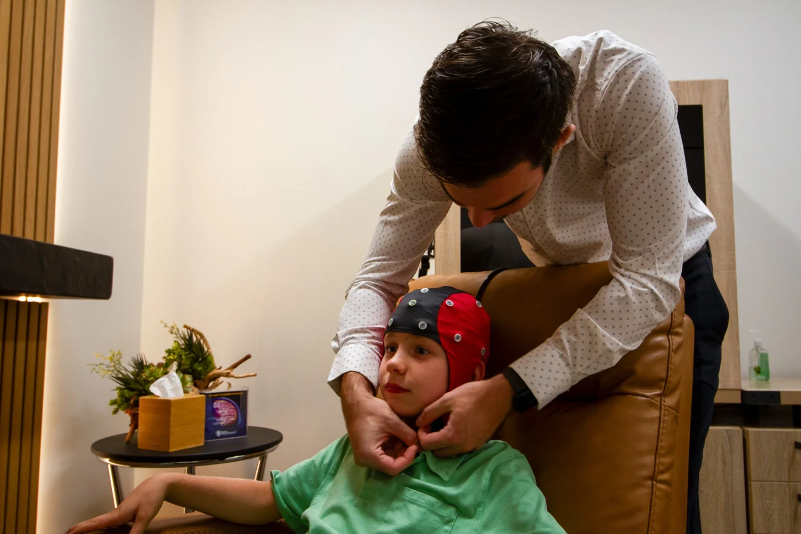
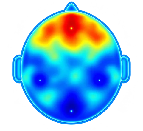
A brain map turns your brain’s electrical activity into a visual picture. Sensors record activity from the scalp, and the results are displayed as color-coded maps that help us examine patterns that may be overactive, underactive, or communicating less efficiently. The recording is safe, non-invasive, and comfortable. We start with objective measurements of your unique brain.
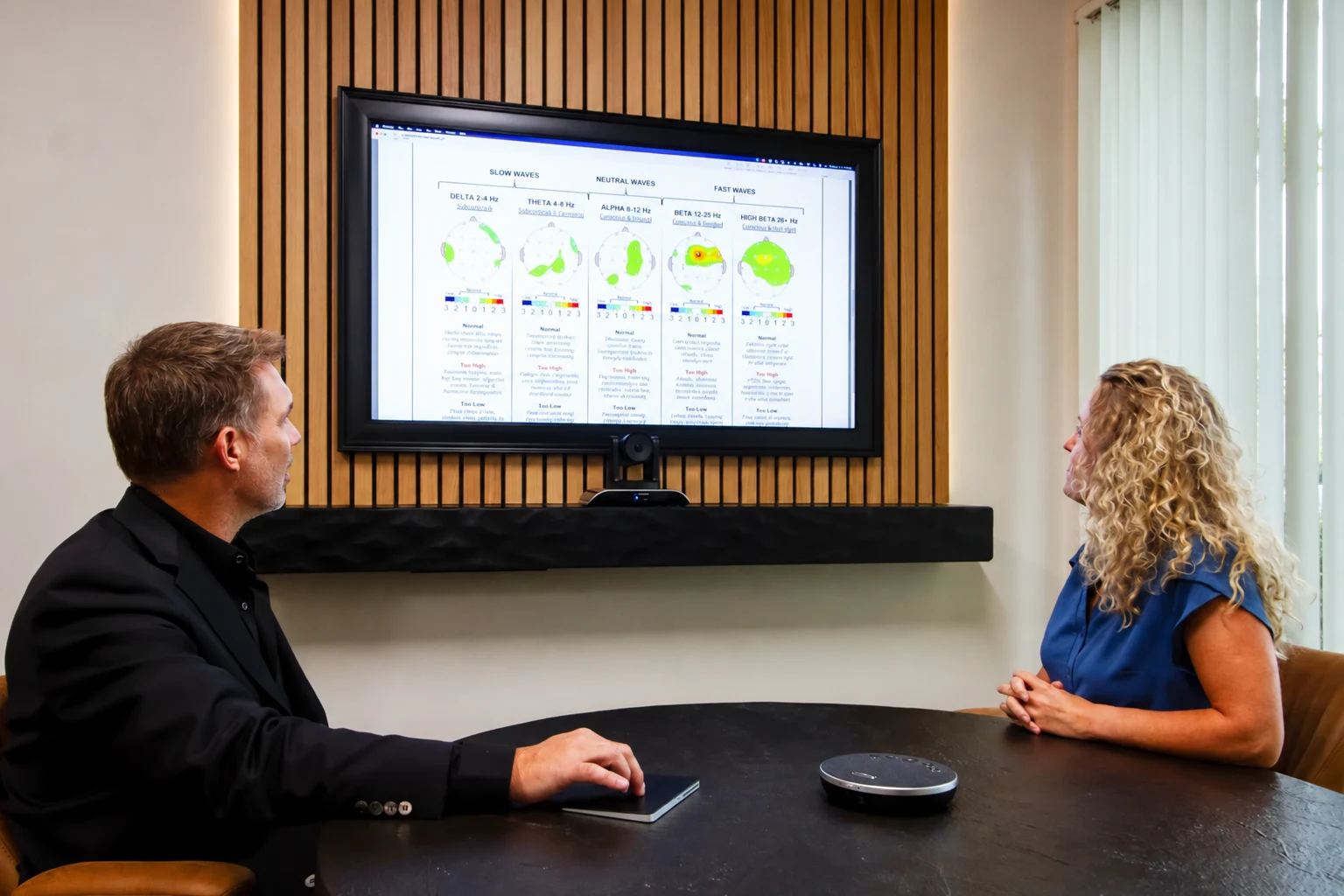
We review your brain map with you and explain which patterns and networks may need support. We also consider sleep, nutrition, genetics, stress, past concussions, and other factors that may affect brain function. Together, this information helps guide a personalized plan for brain training and other appropriate support.
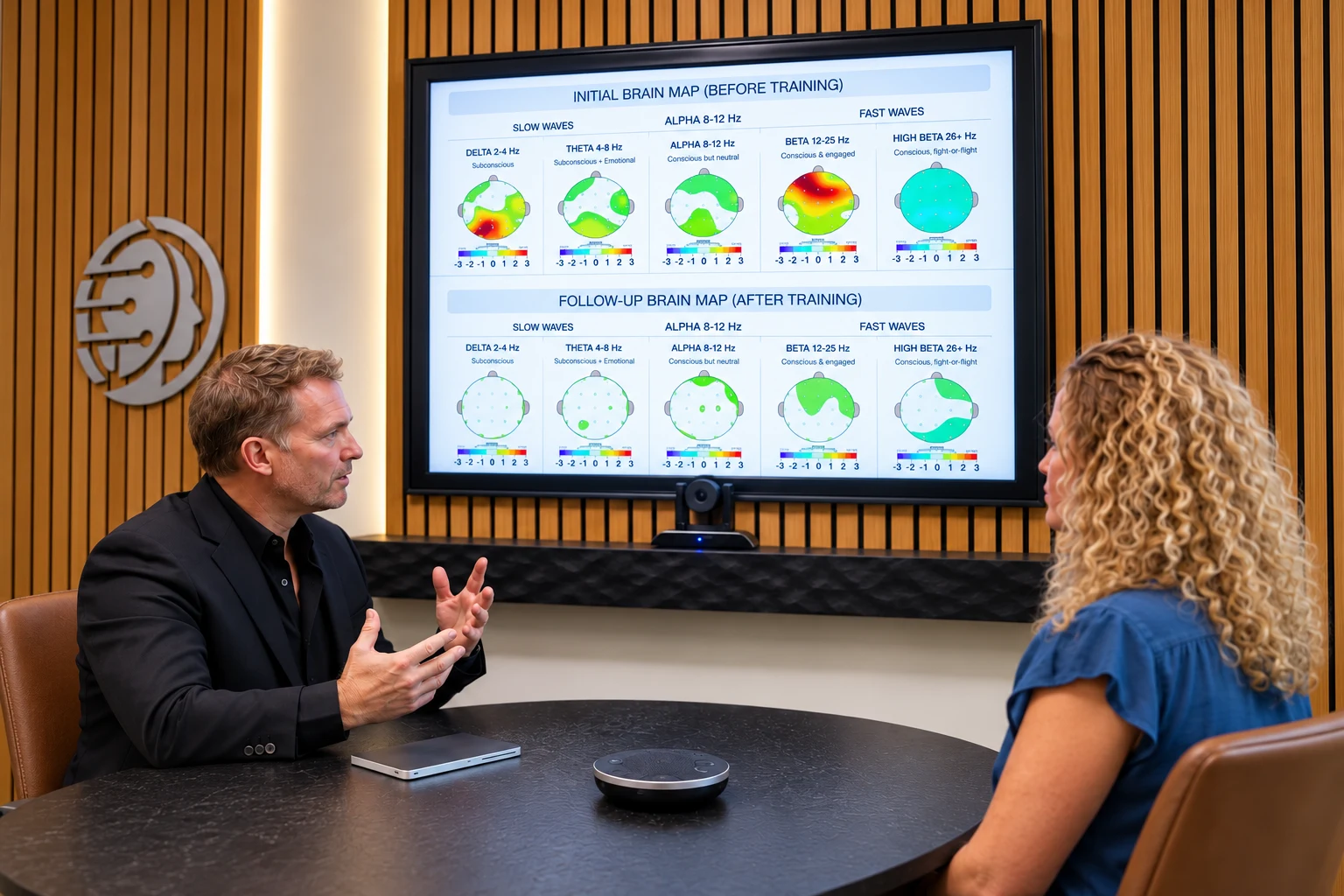
Throughout your program, we repeat your brain map and compare it with your starting point. This helps us see how patterns of brain activity are changing, identify areas of progress, and adjust your plan when additional support is needed. We consider these measurements alongside how you feel and function in daily life.
Explore the research
Select a topic to explore research on measurable EEG patterns.
Distinct EEG patterns within ADHD.
See the research →Resting rhythms and arousal patterns in panic disorder.
See the research →Alpha activity in networks involved in visual processing.
See the research →Regional brainwave differences associated with trauma.
See the research →Quantitative trends during continuous hospital EEG monitoring.
See the research →Measurable brainwave patterns between migraine attacks.
See the research →Fast-frequency activity measured during sleep.
See the research →Coordination of electrical activity across brain regions.
See the research →